Specifications
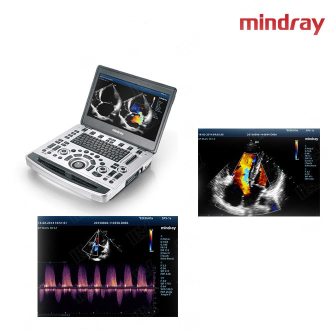
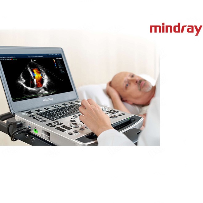
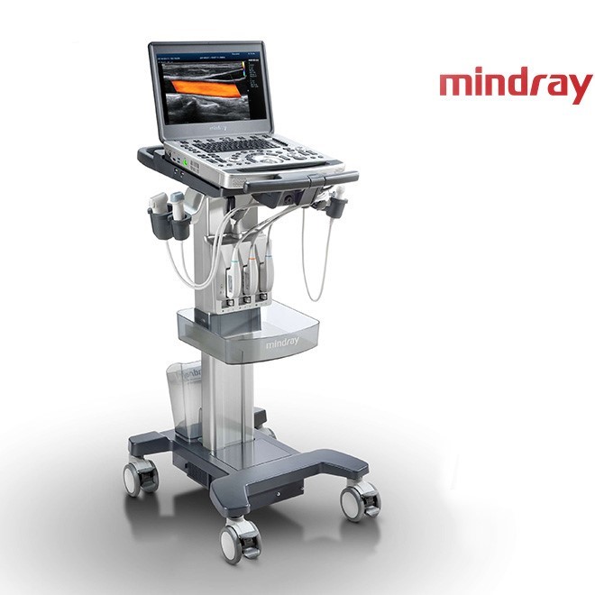
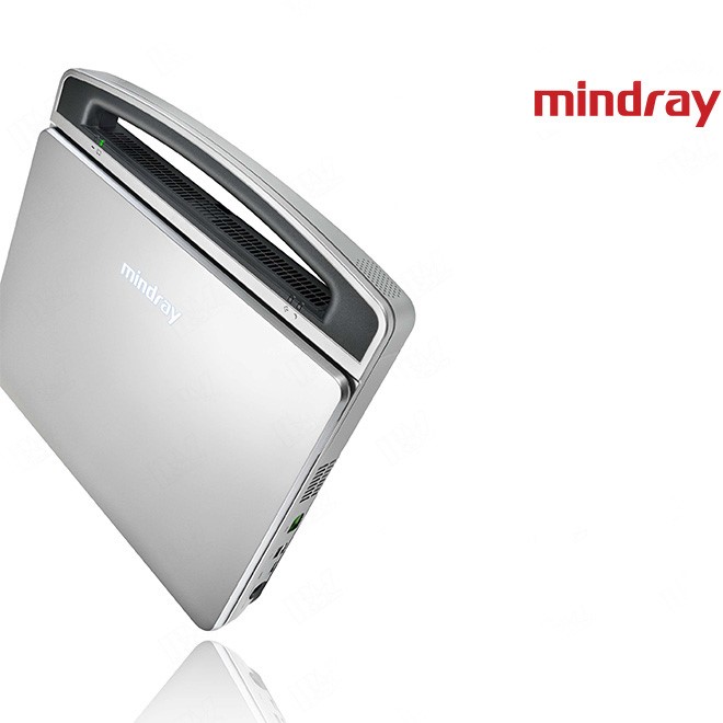
Based on Mindray’s new generation ultrasound platform, mQuadro, M9 has raised the industry standards to an all new level. Advanced signal transmission and reception processors provide highly sensitive and accurate echo detection. Innovative transducer technologies allow for better penetration, higher resolution, greatly enhancing your diagnostic experience.
Mindray’s unique adaptive signal processing technology with intelligent echo detection, designed to utilize the native signal-to-noise information to enhance the weak echo signals while suppressing the surrounding clutter noise, providing more balanced image brightness and improved visualization of myocardium tissue layers.
Tissue Tracking with Quantitative Analysis
The TT QA functionality on M9 allows for a simple, quick and non-invasive solution for the evaluation of left ventricular wall motion abnormalities. Supported by Mindray’s patented 3T technology with single crystal, M9 significantly improves the tracking accuracy and effectiveness, controlling the image drift caused by the probe movement and respiration. With the added unique benefit of on-site analyses, the TT QA on M9 can be performed at the bed side, saving time and making complicated diagnoses much simpler.
LVO with Stress Echocardiography
M9’s premium capabilities allow for LV opacification during stress, enhancing discrimination between myocardial tissue and blood pool, providing better visualization of the endocardial surface. Stress Echo feature on M9 includes a complete package for pharmacological stress and exercise stress echo. The package is supported by a flexible reporting system that can be optimized for your individualistic needs.
PSHITM (Phase Shift Harmonic Imaging)
Purified Harmonic Imaging for better contrast resolution providing clearer images with excellent resolution and less noise
Tissue Harmonic Imaging (THI)
Utilizing second harmonics generated from tissue boundary layers, THI significantly enhances contrast resolution and improves image quality especially for technically difficult subjects.
Tissue Specific Imaging (TSI)
Tissue Specific Imaging optimizes the image quality based on the properties of the tissue being scanned. Four imaging options are available including general, muscle, fluid and fat.
iBeamTM
Permits use of multiple scanned angles to form a single image, resulting in enhanced contrast resolution and improved visualization
iClearTM
Gain improved image quality based on auto structure detection
