4D Color Doppler Ultrasound System NEUCU41
- Advanced 4D Color Doppler Ultrasound System for precise imaging.
- Dual monitors: 19’’ medical monitor and 10.4’’ touch monitor.
- Versatile imaging modes including 2D, 3D, 4D, and various Doppler options.
- Features like Automatic Image Optimization and Tissue Harmonics Imaging for enhanced diagnostics.
- DICOM compliant with export options: USB, Ethernet, and various image formats.
- Supports multiple applications: Abdominal, OB/GYN, Cardiac, Urology, and more.
- Real-time three synchronous unit displaying 2D and Doppler simultaneously.
- High resolution and sensitivity with a dynamic range of up to 260 dB.
- Customizable user interface with preset parameters for efficiency.
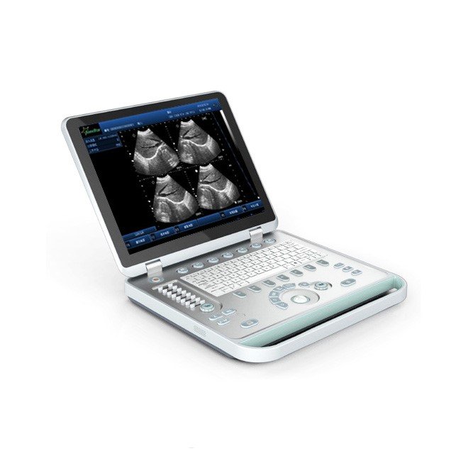
- Lightweight and portable design with various probe options available.
- SKU
- NEUCU41
- Brand
- MedGroup
- Availability
- In stock
- ✓ Warranty included
- ✓ Worldwide shipping


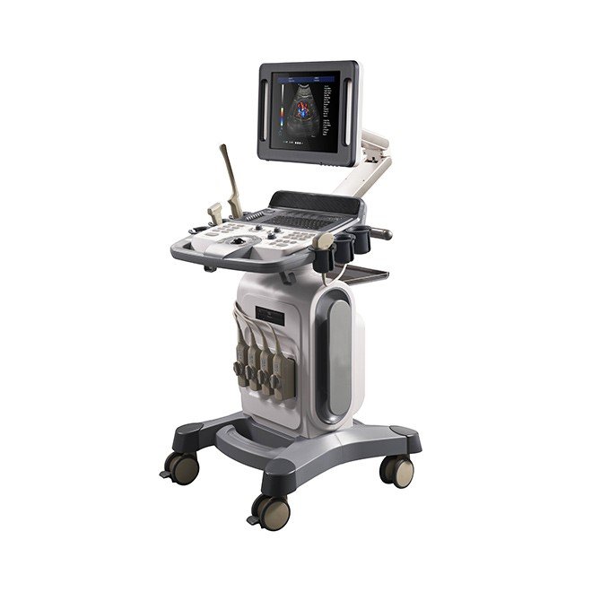
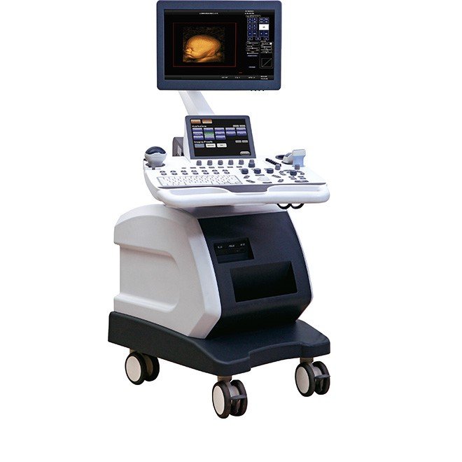
4D Color Doppler Ultrasound System NEUCU41
Advanced multi-application diagnostic imaging system with real-time 4D technology, dual monitors, and comprehensive Doppler capabilities. Delivers exceptional image quality across abdominal, cardiac, OB/GYN, vascular, and musculoskeletal applications with intelligent automation and DICOM compliance.


Clinical Overview
The 4D Color Doppler Ultrasound System NEUCU41 represents a breakthrough in diagnostic imaging technology, delivering exceptional diagnostic capabilities across multiple clinical specialties. Built on a Windows Embedded operating system with Intel i5 processor and dual-monitor configuration, this advanced system combines a 19-inch medical-grade monitor with a 10.4-inch touch screen interface for intuitive operation and optimal visualization. The system's comprehensive imaging modes include 2D, 3D, 4D, Color Doppler, Power Doppler, PW/CW Doppler, and Tissue Doppler, providing healthcare professionals with complete diagnostic flexibility for complex clinical assessments.
Engineered for clinical excellence, the NEUCU41 features advanced technologies including Compound Imaging, Speckle Reduction Imaging, and Tissue Harmonics Imaging that enhance image quality and diagnostic accuracy. The system supports four active probe ports with a wide range of transducer types including convex, linear, sector phased, micro convex, 4D volume convex, endocavity, and veterinary linear probes. With DICOM compliance and multiple export options including USB, Ethernet, and network storage, the NEUCU41 seamlessly integrates into modern healthcare IT infrastructures while delivering superior imaging performance for abdominal, OB/GYN, cardiac, vascular, small parts, pediatric, and musculoskeletal applications.
Key Features & Surgeon Benefits
Superior Resolution & Clarity
- Dynamic range up to 260 dB with 15–145 dB visible adjustment
- Longitudinal resolution ≤1 mm, horizontal ≤0.5 mm
- Scanning depth ≥360 mm for comprehensive penetration
- Compound imaging reduces artifacts and enhances border definition
- Tissue harmonic imaging with three advanced technologies
Intelligent Automation & Efficiency
- One-key automatic optimization adjusts all parameters instantly
- Customizable presets for liver, kidney, gallbladder, uterus, ovaries
- Five programmable shortcut buttons for frequent functions
- Dual-monitor configuration separates imaging from control
- Task lamp navigation guides operators through procedures
Multi-Specialty Capability
- Eight primary clinical applications with specialized measurement packages
- Four active probe ports supporting seven transducer types
- Real-time 4D imaging for obstetric and cardiac applications
- Full Doppler suite: Color, Power, PW, CW, Tissue, Directional
- Supports abdominal, cardiac, OB/GYN, vascular, urology, pediatric, and musculoskeletal
Seamless Data Management
- Full DICOM 3.0 compliance with Store, Print, Working List, Structured Reports
- Multiple export formats: DICOM, JPG, BMP, PNG, AVI
- 560GB storage capacity with E-COM graphic management system
- Ethernet, USB, DVD/CD, and network storage connectivity
- Editable bilingual diagnostic reports with embedded images
Technical Specifications
| Model | MSLCU41 3.0 VERSION |
|---|---|
| SKU | NEUCU41 |
| Operating System | Windows Embedded (CN, EN language) |
| Processor | Intel i5 with 4GB RAM, 120GB SSD + 500GB HDD |
| Display | 19" medical monitor (1280×1024) + 10.4" LED touch screen |
| Imaging Modes | 2D, 3D, 4D, Color Doppler, Power Doppler, PW/CW Doppler, Tissue Doppler, Color M-Mode, Free Steering M-Mode |
| Advanced Features | Compound Imaging, Speckle Reduction, Tissue Harmonics, 4D Real-Time, Auto Optimization, Tissue Doppler, Multi-Beam, IMT, Trapezoidal Imaging, iBank Database |
| Dynamic Range | Up to 260 dB (15–145 dB visible adjustable) |
| Image Resolution | Longitudinal ≤1 mm, Horizontal ≤0.5 mm |
| Scanning Depth | ≥360 mm |
| Probe Ports | 4 Active Ports |
| Transducer Types | Convex (2.5–5.0 MHz), Linear (6.0–14.0 MHz), Sector Phased (2.0–5.5 MHz), Micro Convex, 4D Volume Convex (2.0–5.5 MHz), Endocavity (5.0–9.0 MHz), Veterinary Linear |
| Blood Flow Sensitivity | ≤1 mm/s |
| Maximum Blood Flow Velocity | PWD ≥12,300 mm/s, CW ≥33,200 mm/s |
| Sample Width Range | 0.5–40 mm |
| Cine Memory | >10 seconds, 750 frames |
| Scanning Angles | Abdominal probe ≥105°, Intracavity probe ≥160°, Convex probe 20°–85° adjustable |
| Storage Capacity | 560GB Hard Disk |
| DICOM Modes | Store, Print, Working List, Storage Commitment, Structured Reports |
| Export Options | DICOM, Ethernet, JPG/BMP/PNG, AVI, Network Storage, USB, DVD/CD+R(W) |
| Input/Output | VGA, 2 USB Ports, Ethernet, DICOM, Built-in Speakers |
| Applications | Abdominal, OB/GYN, Urology, Cardiac, Vascular, Small Parts, Pediatric, Musculoskeletal |
| Warranty | 2 Year Manufacturer Warranty |
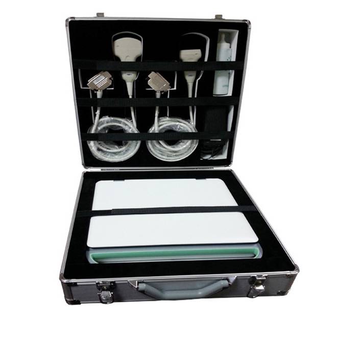
Standard Instrument Composition
| Item | Description | Qty |
|---|---|---|
| Main Console Unit | NEUCU41 ultrasound system with Windows Embedded OS, Intel i5 processor, dual monitors | 1 |
| 19" Medical Monitor | High-resolution medical-grade display (1280×1024) | 1 |
| 10.4" Touch Screen Monitor | LED touch interface for menu and parameter control | 1 |
| Probe Ports | Four active transducer connection ports | 4 |
| Convex Probe | 2.5–5.0 MHz for abdominal imaging | 1 |
| Linear Probe | 6.0–14.0 MHz for small parts and musculoskeletal | 1 |
| Phased Array Probe | 2.0–5.5 MHz for cardiac imaging | 1 |
| 4D Volume Probe | 2.0–5.5 MHz for real-time 4D obstetric imaging | 1 |
| Endocavity Probe | 5.0–9.0 MHz for transvaginal examinations | Optional |
| Micro Convex Probe | Specialized convex design for pediatric applications | Optional |
| Veterinary Linear Probe | Specialized probe for veterinary applications | Optional |
| Gel Warmer | Maintains optimal ultrasound gel temperature | 1 |
| Probe Holders | Secure mounting for transducers | 4 |
| Power Supply | AC power adapter and cable | 1 |
| USB Connectivity | 2 USB ports for data export and peripherals | 2 |
| Ethernet Cable | For DICOM and network connectivity | 1 |
| Documentation | User manual, technical specifications, warranty certificate | 1 set |
Get in Touch for Pricing & Availability
The NEUCU41 is a premium diagnostic ultrasound system designed for multi-specialty imaging centers, hospitals, and advanced clinical practices. Contact our sales team for detailed pricing, lease options, probe configurations, and volume discounts.
Available on request
Configuration, paperwork & clinical fit
The full spec + documentation pack for this device ships ahead of purchase on request. A sales engineer responds within one business day.
Configuration sheet
Specifications, accessories & clinical fit
Sizes and dimensions, material and finish, compatible instruments and screw references, what ships in the set, lead time and warranty — all in one specification sheet, sent within one business day.
Documents & downloads
Regulatory paperwork & manuals
This device's IFU, CE Declaration of Conformity, ISO 13485 certificate and (where applicable) FDA 510(k) clearance ship with the order. Reach out below for the pack ahead of purchase.
Get in touch
Need configuration help, certificates, or volume pricing?
Our clinical sales team will scope quantities, voltage, accessories and regulatory paperwork — usually within one business day.
Customer reviews
No reviews yet — be the first to share your experience.
Frequently bought together
Related products
See all →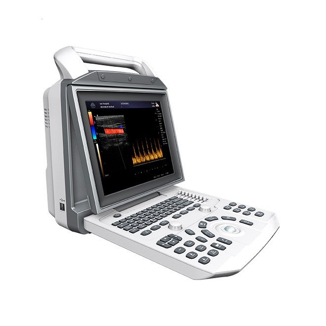
Full Digital Color Doppler Ultrasonic Diagnostic System NEUCU45M
- High-definition 12.1″ LCD monitor with adjustable viewing angle - Dual transducer ports for versatile imaging capabilities - Long-lasting lithium battery for over 2 hours of operation - Advanced imaging technologies including speckle noise removal and tissue harmonic imaging - Comprehensive software packages for various medical uses: Abdomen, Cardiac, Obstetrics, and more - DICOM 3.0 compatibility for seamless data sharing - Multiple data interface options: USB2.0, VGA, and more - Lightweight design: Net weight of 8KG, gross weight of 14KG - Extensive clinical applications across various medical disciplines - Unique puncture guide and automated measurement technologies for enhanced accuracy

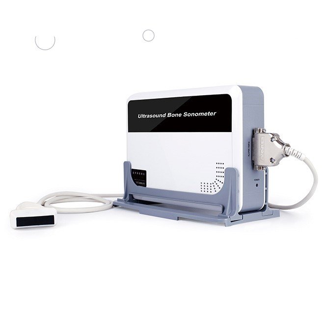
Portable Bone Density Scanner NEUBD10
- Portable and lightweight design for easy transport - Utilizes ultrasound technology for non-invasive bone density measurement - Measures bone density at the radius and tibia, suitable for all ages - Ideal for pregnant women, children, and the elderly - Provides reliable results unaffected by soft tissue thickness or bone size - Quick measurement time: single measurement in ≤ 25 seconds - Compatible with Windows XP and above operating systems - Features a comprehensive database for multi-age data selection - Operating conditions: 5-40℃ and relative humidity ≤ 80% - Easy to connect to desktop computers, laptops, or tablets

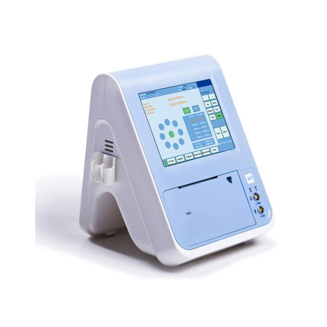
A B Scan Ophthalmic Ultrasound Pachymeter NEUPU21
- 5.7 inch color touch LCD screen for easy navigation - Rechargeable battery lasting up to 10 hours - Built-in thermal printer with one-key print function - USB port for direct PC connectivity - Supports I-station workstation connection - Lightweight and portable design at 1.7Kg - Multi-language support: Chinese, English, Russian, French, Spanish, and Portuguese - High-resolution measurement accuracy of ±0.02mm - Examination modes include normal, aphakic, pseudophakic, and dense cataract - Versatile measurement modes: immersion, contact, auto, and manual

3D Ultrasound Machine NEUCU23
- 10.4" color LCD full screen display with adjustable angle - One-key-light keyboard for use in dark environments - Easy one-button operation for storage, review, and printing of images - Quick access to stored images for enhanced efficiency - Multi-functional knob allowing quick adjustments across modes - Advanced imaging techniques for optimized signal accuracy - Adaptive color artifact removal for clear images - Linear probe with independent angle deflection - Integrated working station for streamlined operation - Abundant measuring software for comprehensive data management
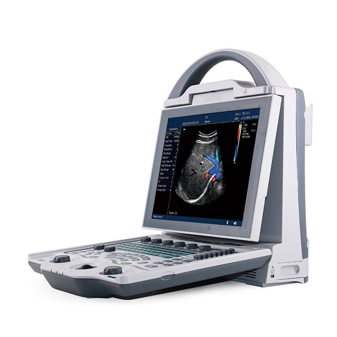
3D Laptop Ultrasound Machine NEUPU34
- 15 inch LCD display for clear imaging - Wide working frequency range: 2.0MHz to 10MHz - Multiple display modes: B, B+B, B+M, M, and 4B - Convex array scanning range of 60° to 150° - Adjustable amplification factor (1.0, 1.2, 1.5, 2.0) - 8 segments TGC and overall gain control - Advanced image processing techniques for enhanced clarity - Supports 512 lines/frame with a 30 fps frame rate - Convenient USB flash disk image storage - Built-in battery allows over 5 hours of operation - Weighs only 4Kg, including battery for portability
3D Full Digital Laptop Ultrasound Scanner NEUPU25
- High-resolution 15 inch LCD display for clear imaging - Versatile working frequency range of 2.0MHz to 10MHz - Multiple display modes: B, B+B, B+M, M, and 4B - Wide scanning range from 60° to 150° with convex array - Advanced image processing with 8 γ corrections and digital edge enhancement - 512 lines/frame scanning for enhanced detail and clarity - Fast frame rate of 30 frames per second for real-time imaging - USB flash disk support for large volume image storage - Built-in battery allowing over 5 hours of continuous use - Lightweight design at only 4Kg, including battery and standard configurations

4D Full Digital Trolley Color Doppler Ultrasound NEUCU30
- 4D Full Digital Trolley Color Doppler Ultrasound NEUCU30 - Supports multiple displaying modes: B, M, Doppler, and more - Advanced full-digital signal processing for superior imaging - Intelligent imaging technologies including THI and iBeam TM - Extensive measurement packages for abdomen, OB, gynecological, urology, cardiac, and vascular assessments - Adjustable parameters such as Doppler sound output, wall filter, and sampling frame - High storage capacity with 500G available for images in BMP, JPEG, and PNG formats - Standard configuration includes a 15-inch LED monitor and dual probes - Optional accessories available for expanded functionality, including transvaginal probes and DICOM 3.0 compatibility.