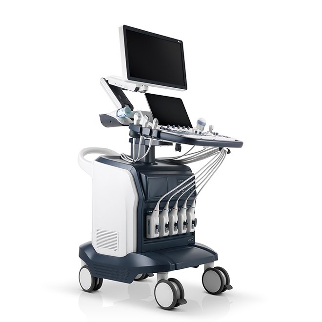
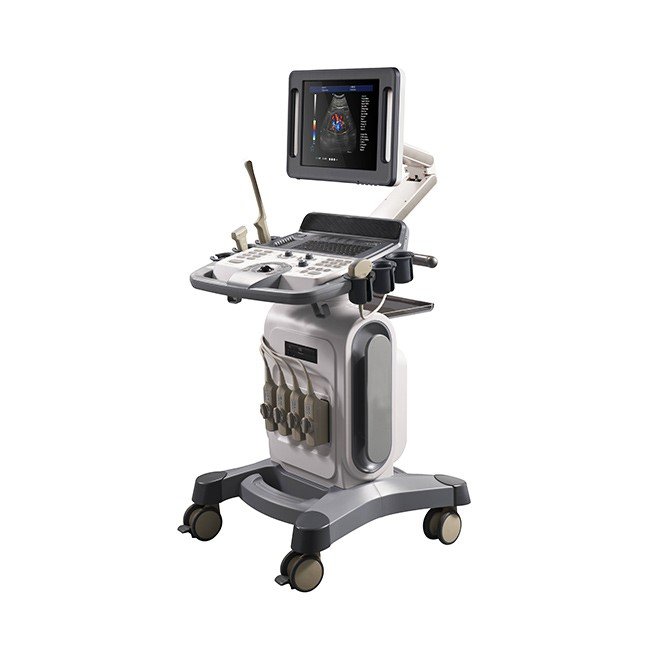
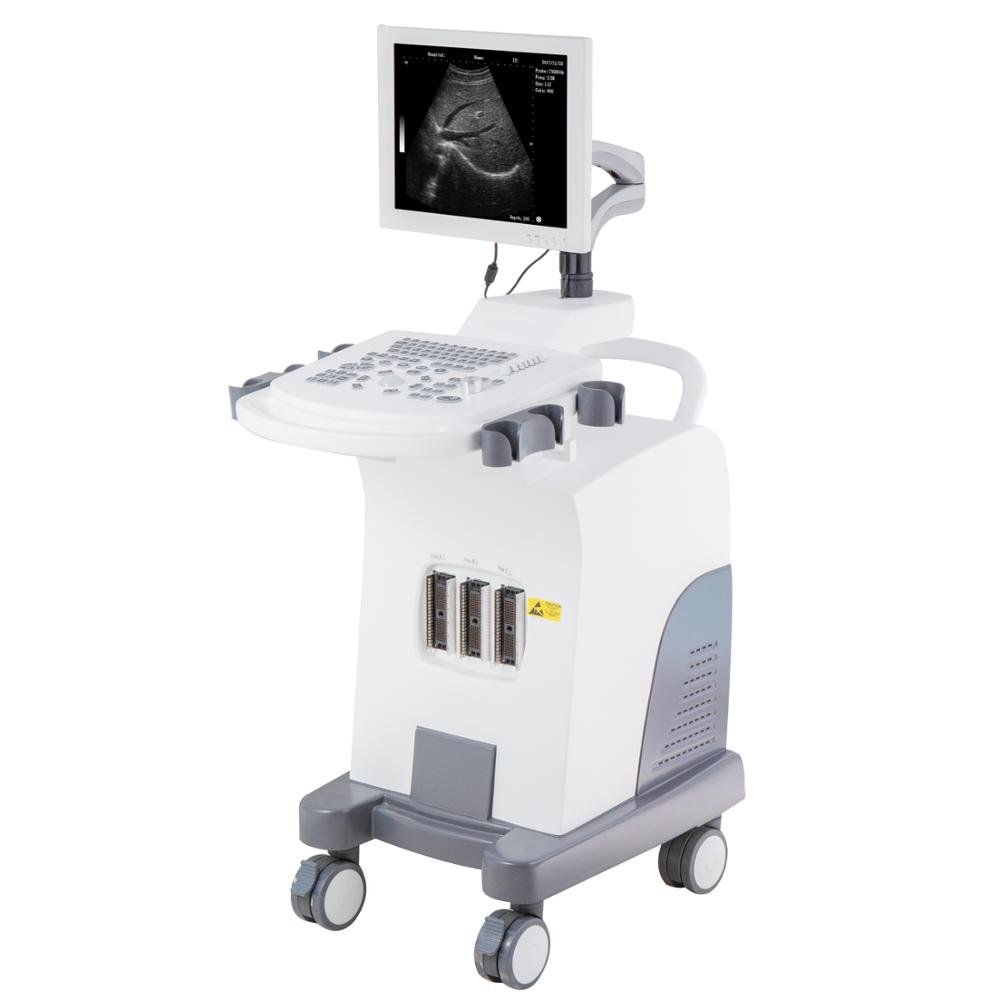
SonoScape S60 Ultrasound Machine
- Advanced SonoScape S60 Ultrasound Machine for versatile applications including obstetrics, cardiology, and musculoskeletal imaging.
- Multiple imaging modes available: B, M, Color M, 3D/4D, and more for comprehensive diagnostics.
- Supports various probes: convex, linear, phased array, and volume for tailored examinations.
- High-resolution 1920*1080 LCD touchscreen with adjustable swivel and viewing angles.
- User-friendly control panel with customizable layout and backlight design for efficient operation.
- Compact dimensions- weighs 123 Kg, conveniently designed for easy movement and storage.
- Equipped with high-quality Hi-Fi speakers for enhanced audio output during procedures.
- Multiple language support for software and user manual including English, Spanish, and Chinese.
- Designed for a wide range of working environments with temperature and humidity tolerance.
- Includes advanced features like 3D/4D imaging, automatic functions, and multi-level controls.
- SKU
- S60
- Brand
- SonoScape
- Availability
- In stock
- ✓ Warranty included
- ✓ Worldwide shipping


SonoScape S60 Ultrasound Machine
Premium diagnostic ultrasound system with advanced 3D/4D imaging, high-resolution 1920×1080 Full HD touchscreen display, and comprehensive clinical capabilities. Professional-grade technology for obstetrics, cardiology, musculoskeletal, vascular, and multi-specialty applications with intelligent automation and complete Doppler suite.


Clinical overview
The SonoScape S60 represents a comprehensive diagnostic ultrasound platform engineered for high-volume clinical environments and specialized applications. With its Full HD 1920×1080 medical-grade touchscreen, advanced 3D/4D imaging capabilities, and complete Doppler suite, the S60 delivers exceptional diagnostic confidence across obstetrics, cardiology, abdominal, musculoskeletal, and vascular imaging. The system's intelligent automation features—including Auto-Trace, Auto-NT, and stress echocardiography—reduce operator variability while accelerating workflow efficiency.
Supporting five probe ports with simultaneous connectivity to convex, linear, phased array, and volume transducers, the S60 eliminates connection delays in multi-specialty practices. Advanced imaging enhancements including compound imaging, trapezoid mode, panoramic view, and tissue harmonic imaging extend diagnostic capabilities beyond conventional B-mode scanning. Contrast-enhanced ultrasound (CEUS) with Time-Intensity Curve analysis, integrated biopsy guidance, and comprehensive Doppler modalities (Color, Power, PW, CW, HPRF) position the S60 as a versatile solution for complex diagnostic and interventional procedures. The ergonomic control panel with customizable layouts, backlit controls, and adjustable trackball sensitivity optimizes operator comfort during extended examination sessions, while 13-language software support ensures accessibility in international healthcare environments.
Key features & surgeon benefits
Premium Full HD Touchscreen
- 1920×1080 resolution medical-grade LCD with 178° viewing angles
- ±60° swivel and −45° to +25° vertical tilt for ergonomic positioning
- Direct touchscreen parameter adjustment for intuitive workflow
- User-customizable interface layouts and backlit controls
3D/4D & Multi-Mode Capabilities
- Real-time 4D imaging for dynamic fetal and cardiac visualization
- Freehand 3D reconstruction without specialized volume probes
- B, M, Color M, PW Doppler, CW Doppler, HPRF, Color Doppler, Power Doppler, Directional Power Doppler
- Compound imaging, trapezoid mode, panoramic view, and tissue harmonic imaging
Intelligent Measurement Tools
- Auto-Trace for automated cardiac boundary detection and measurement
- Auto-NT for first-trimester nuchal translucency screening
- Stress echocardiography with multi-stage acquisition and wall motion scoring
- Automated obstetric biometry with fetal growth percentile calculations
Multi-Probe & DICOM Support
- Five probe ports (four active) for instant transducer switching
- Full DICOM 3.0 integration with PACS and HIS/RIS systems
- Modality Worklist (MWL) for automated patient demographics import
- 13-language software support for global clinical environments
Technical specifications
| Model | SonoScape S60 |
|---|---|
| Display | 1920×1080 LCD Medical-Grade Touchscreen, 178° Viewing Angles |
| Display Adjustment | ±60° Swivel, −45° to +25° Vertical Tilt |
| Imaging Modes | B, M, Color M, PW Doppler, CW Doppler, HPRF, Color Doppler, Power Doppler, Directional Power Doppler, 3D/4D |
| Advanced Features | Compound Imaging, Trapezoid Imaging, Panoramic View, Auto-Trace, Auto-NT, Stress Echo, Contrast Imaging (TIC), Biopsy Guidance |
| Probe Ports | Five Ports (Four Active/Interchangeable, One Inactive) |
| Supported Probes | Convex (2–6 MHz), Linear (5–14 MHz), Phased Array (2–4 MHz), Endocavity, Pediatric, 3D/4D Volume |
| TGC Controls | Eight-Level Slider System |
| Trackball Sensitivity | Adjustable (0–150 mm Range) |
| Frequency Control | 5-Band Adjustable (Fundamental and Harmonic Waves) |
| Audio System | Hi-Fi Speaker for Doppler Audio Output |
| Dimensions (W×D×H) | 573 mm × 982 mm × 1344 mm |
| Weight | 123 kg |
| Power Input | 100–110V/240V~, 50/60 Hz |
| Maximum Power Output | 300 W |
| Operating Temperature | 0–40°C |
| Humidity Tolerance | 30–85% RH (Non-Condensing) |
| System Noise Level | ≤55 dB |
| Software Languages | 13 Languages Supported |
| Certifications | FDA, CE, ISO 13485 |
| DICOM Support | Full DICOM 3.0 with PACS, HIS/RIS Integration, Modality Worklist (MWL) |
| Warranty | 2 Year Manufacturer Warranty |
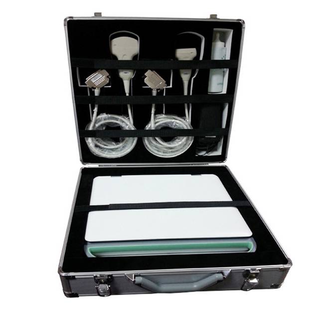
Standard instrument composition
| Item | Description | Qty |
|---|---|---|
| Main Console Unit | SonoScape S60 Ultrasound System with Integrated Cart and Casters | 1 |
| Display Monitor | 1920×1080 Full HD LCD Touchscreen with Swivel Mount | 1 |
| Control Panel | User-Customizable Panel with Backlit Controls and Trackball | 1 |
| Power Supply | 100–110V/240V AC, 50/60 Hz Input Module | 1 |
| Probe Cables | Five Probe Port Connectors (Four Active, One Inactive) | 5 |
| Audio System | Integrated Hi-Fi Speaker for Doppler Output | 1 |
| Documentation | User Manual, Technical Specifications, Safety Guidelines (13 Languages) | 1 Set |
| Warranty Card | 2-Year Manufacturer Warranty Documentation | 1 |
Get in touch for pricing & availability
Contact our sales team to discuss the SonoScape S60 specifications, probe options, financing programs, and on-site demonstration scheduling. We provide comprehensive training and ongoing technical support for all installations.
Email: [email protected]
Hours: Monday–Friday, 8 AM–6 PM PST | 24/7 Emergency Support Available
Available on request
Configuration, paperwork & clinical fit
The full spec + documentation pack for this device ships ahead of purchase on request. A sales engineer responds within one business day.
Configuration sheet
Specifications, accessories & clinical fit
Sizes and dimensions, material and finish, compatible instruments and screw references, what ships in the set, lead time and warranty — all in one specification sheet, sent within one business day.
Documents & downloads
Regulatory paperwork & manuals
This device's IFU, CE Declaration of Conformity, ISO 13485 certificate and (where applicable) FDA 510(k) clearance ship with the order. Reach out below for the pack ahead of purchase.
Get in touch
Need configuration help, certificates, or volume pricing?
Our clinical sales team will scope quantities, voltage, accessories and regulatory paperwork — usually within one business day.
Customer reviews
No reviews yet — be the first to share your experience.
Frequently bought together
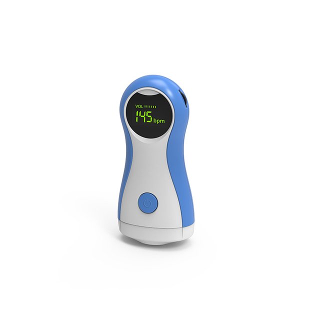
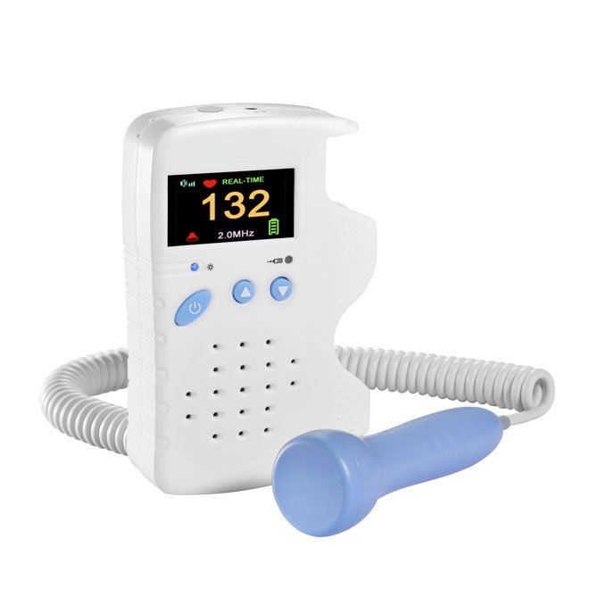
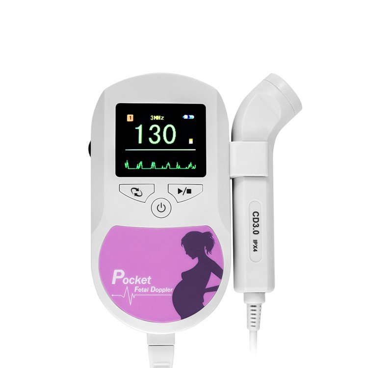
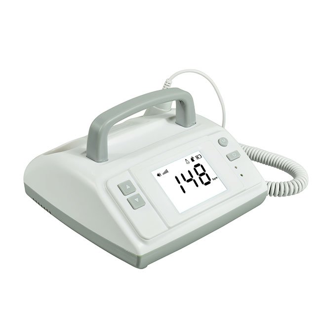
Related products
See all →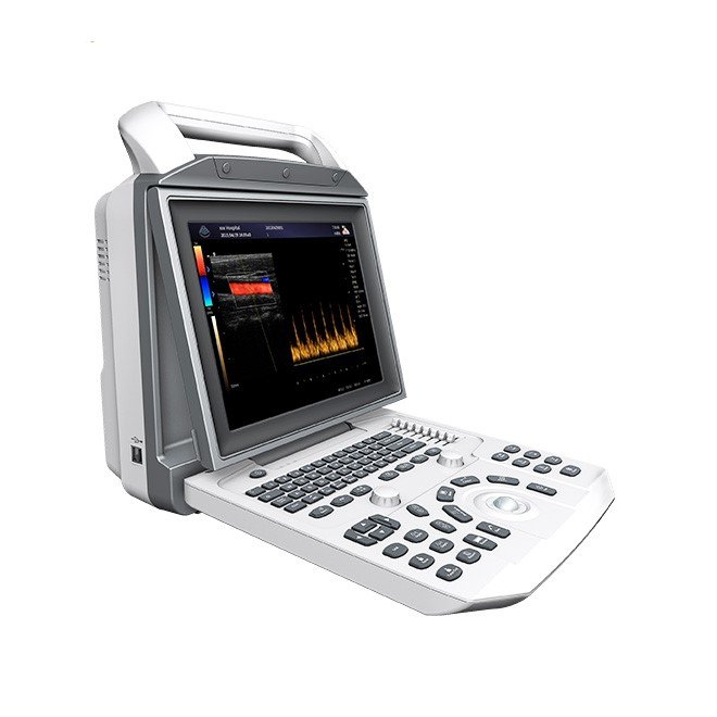
Full Digital Color Doppler Ultrasonic Diagnostic System NEUCU45M
- High-definition 12.1″ LCD monitor with adjustable viewing angle - Dual transducer ports for versatile imaging capabilities - Long-lasting lithium battery for over 2 hours of operation - Advanced imaging technologies including speckle noise removal and tissue harmonic imaging - Comprehensive software packages for various medical uses: Abdomen, Cardiac, Obstetrics, and more - DICOM 3.0 compatibility for seamless data sharing - Multiple data interface options: USB2.0, VGA, and more - Lightweight design: Net weight of 8KG, gross weight of 14KG - Extensive clinical applications across various medical disciplines - Unique puncture guide and automated measurement technologies for enhanced accuracy

Portable Bone Density Scanner NEUBD10
- Portable and lightweight design for easy transport - Utilizes ultrasound technology for non-invasive bone density measurement - Measures bone density at the radius and tibia, suitable for all ages - Ideal for pregnant women, children, and the elderly - Provides reliable results unaffected by soft tissue thickness or bone size - Quick measurement time: single measurement in ≤ 25 seconds - Compatible with Windows XP and above operating systems - Features a comprehensive database for multi-age data selection - Operating conditions: 5-40℃ and relative humidity ≤ 80% - Easy to connect to desktop computers, laptops, or tablets

A B Scan Ophthalmic Ultrasound Pachymeter NEUPU21
- 5.7 inch color touch LCD screen for easy navigation - Rechargeable battery lasting up to 10 hours - Built-in thermal printer with one-key print function - USB port for direct PC connectivity - Supports I-station workstation connection - Lightweight and portable design at 1.7Kg - Multi-language support: Chinese, English, Russian, French, Spanish, and Portuguese - High-resolution measurement accuracy of ±0.02mm - Examination modes include normal, aphakic, pseudophakic, and dense cataract - Versatile measurement modes: immersion, contact, auto, and manual

3D Ultrasound Machine NEUCU23
- 10.4" color LCD full screen display with adjustable angle - One-key-light keyboard for use in dark environments - Easy one-button operation for storage, review, and printing of images - Quick access to stored images for enhanced efficiency - Multi-functional knob allowing quick adjustments across modes - Advanced imaging techniques for optimized signal accuracy - Adaptive color artifact removal for clear images - Linear probe with independent angle deflection - Integrated working station for streamlined operation - Abundant measuring software for comprehensive data management
3D Laptop Ultrasound Machine NEUPU34
- 15 inch LCD display for clear imaging - Wide working frequency range: 2.0MHz to 10MHz - Multiple display modes: B, B+B, B+M, M, and 4B - Convex array scanning range of 60° to 150° - Adjustable amplification factor (1.0, 1.2, 1.5, 2.0) - 8 segments TGC and overall gain control - Advanced image processing techniques for enhanced clarity - Supports 512 lines/frame with a 30 fps frame rate - Convenient USB flash disk image storage - Built-in battery allows over 5 hours of operation - Weighs only 4Kg, including battery for portability
3D Full Digital Laptop Ultrasound Scanner NEUPU25
- High-resolution 15 inch LCD display for clear imaging - Versatile working frequency range of 2.0MHz to 10MHz - Multiple display modes: B, B+B, B+M, M, and 4B - Wide scanning range from 60° to 150° with convex array - Advanced image processing with 8 γ corrections and digital edge enhancement - 512 lines/frame scanning for enhanced detail and clarity - Fast frame rate of 30 frames per second for real-time imaging - USB flash disk support for large volume image storage - Built-in battery allowing over 5 hours of continuous use - Lightweight design at only 4Kg, including battery and standard configurations

4D Full Digital Trolley Color Doppler Ultrasound NEUCU30
- 4D Full Digital Trolley Color Doppler Ultrasound NEUCU30 - Supports multiple displaying modes: B, M, Doppler, and more - Advanced full-digital signal processing for superior imaging - Intelligent imaging technologies including THI and iBeam TM - Extensive measurement packages for abdomen, OB, gynecological, urology, cardiac, and vascular assessments - Adjustable parameters such as Doppler sound output, wall filter, and sampling frame - High storage capacity with 500G available for images in BMP, JPEG, and PNG formats - Standard configuration includes a 15-inch LED monitor and dual probes - Optional accessories available for expanded functionality, including transvaginal probes and DICOM 3.0 compatibility.