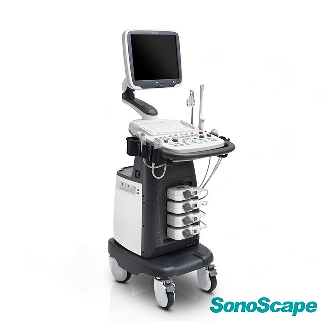
Ultrasound SonoScape S12
– High-resolution 15″ LED color monitor for clear imaging
– Versatile imaging modes: B, 2B, 4B, M, THI, CFM, and more
– Equipped with four transducer connectors for flexibility
– Supports advanced features like Pulse Inversion Harmonic and Compound Imaging
– Includes ECG module for comprehensive diagnostics
– 500GB hard disk storage with options for 1TB upgrades
– Multi-beam technology enhances image clarity
– Easy image optimization with M-Tuning one-button feature
– DICOM 3.0 compatibility for seamless data management
– Freehand 3D Imaging capability for detailed assessments
Ultrasound SonoScape S12
Advanced Diagnostic Imaging with Comprehensive Clinical Applications
The SonoScape S12 ultrasound system delivers exceptional imaging performance with its high-resolution 15-inch LED color monitor, versatile imaging modes including B, 2B, 4B, M, THI, and CFM, and advanced features like Pulse Inversion Harmonic and Compound Imaging. Equipped with four transducer connectors, ECG module, and 500GB storage, this system provides comprehensive diagnostic capabilities for abdominal, vascular, obstetric, and musculoskeletal applications.
ISO Certified
Fast Delivery
Expert Support
Warranty Included
Introduction to the SonoScape S12 Ultrasound System
The SonoScape S12 represents a significant advancement in diagnostic ultrasound technology, combining exceptional image quality with comprehensive clinical functionality. Designed for modern healthcare facilities, this versatile system delivers outstanding performance across multiple medical specialties including radiology, cardiology, obstetrics, gynecology, vascular imaging, and musculoskeletal diagnostics. With its intuitive interface and advanced imaging technologies, the S12 enables clinicians to perform accurate diagnoses with confidence and efficiency.
Featuring a high-resolution 15-inch LED color monitor, the S12 provides crisp, detailed images that enhance diagnostic accuracy. The system comes equipped with four transducer connectors, allowing for quick switching between different examination types without workflow interruption. Standard configuration includes two premium transducers: a 128-element linear array L741 for vascular and small parts imaging, and a 128-element convex array C344 for abdominal and obstetric applications. Advanced software features including Compound Imaging, Pulse Inversion Harmonic, μ-Scan speckle reduction, and Freehand 3D Imaging provide comprehensive diagnostic capabilities in a single, compact platform.
Clinical Applications
Technical Specifications
| Component | Specification |
|---|---|
| Display Monitor | 15-inch High Resolution LED Color Monitor |
| Transducer Connectors | Four Active Transducer Ports |
| Storage Capacity | 500GB Hard Disk (1TB Upgrade Available) |
| Data Management | DVD-RW, USB 2.0, Hard Disk |
| ECG Integration | Built-in ECG Module |
| Imaging Modes | B, 2B, 4B, M, THI, CFM, PDI, DirPDI, PW, HPRF |
| Advanced Imaging | Compound Imaging, Pulse Inversion Harmonic, Trapezoidal Imaging |
| Image Optimization | μ-Scan Speckle Reduction, Multi-beam Technology, M-Tuning |
| 3D Capabilities | Freehand 3D Imaging |
| Measurements | IMT Measurement, Comprehensive Calculation Packages |
| Connectivity | DICOM 3.0 (Store/C-Store/Worklist/MPPS/Print/Q/R) |
| Linear Array Transducer | L741: 128 elements, 4-16MHz, 46mm footprint |
| Convex Array Transducer | C344: 128 elements, 2-6.8MHz, R40mm radius |
| Applications | Vascular, Small Parts, MSK, Abdominal, OB/GYN |
| Gain Compensation | Lateral Gain Compensation (LGC) |
| Needle Guidance | VIS-Needle Enhanced Visualization |
Key Features and Benefits
High-Resolution 15-Inch LED Monitor
The large, high-resolution LED color monitor provides exceptional image clarity with superior brightness and contrast. The 15-inch display offers ample viewing area for detailed examination and comparison of multiple imaging windows simultaneously, reducing eye strain during extended scanning sessions.
Comprehensive Imaging Modes
With B-mode, 2B, 4B, M-mode, Tissue Harmonic Imaging, Color Flow Mapping, Power Doppler Imaging, Directional Power Doppler, Pulsed Wave, and High PRF modes, the S12 provides complete diagnostic versatility for all clinical applications and patient types.
Four Transducer Connectors
Multiple active transducer ports enable seamless workflow transitions between different examination types. Keep your most frequently used transducers connected and ready, eliminating connection delays and maximizing scanning efficiency throughout the day.
Pulse Inversion Harmonic Imaging
Advanced harmonic imaging technology significantly improves image quality by reducing artifacts and enhancing contrast resolution. Tissue Harmonic Imaging provides clearer visualization of difficult-to-image patients, particularly those with challenging body habitus.
Compound Imaging Technology
Spatial compound imaging combines multiple angle views to reduce speckle, enhance borders, and improve visualization of tissue structures. This technology provides more uniform images with reduced artifacts and improved diagnostic confidence.
Freehand 3D Imaging
Three-dimensional imaging capability allows for volumetric data acquisition and reconstruction, providing unique perspectives for complex anatomical assessment. Particularly valuable in obstetric applications for fetal evaluation and patient counseling.
μ-Scan Speckle Reduction
Proprietary 2D speckle reduction technology enhances image quality by reducing acoustic noise while preserving tissue detail. μ-Scan processing results in cleaner, more readable images with improved border definition and tissue differentiation.
Multi-Beam Technology
Advanced multi-beam processing increases frame rates and improves temporal resolution for enhanced visualization of moving structures. This technology ensures smooth, real-time imaging even during demanding cardiac and vascular studies.
VIS-Needle Enhancement
Specialized needle visualization technology dramatically improves needle conspicuity during interventional procedures. VIS-Needle provides clearer needle tracking for safer, more accurate biopsies, aspirations, and guided injections.
M-Tuning One-Button Optimization
Intelligent image optimization automatically adjusts multiple parameters with a single button press. M-Tuning reduces the learning curve for new operators while ensuring consistent, high-quality images across all users and applications.
Integrated ECG Module
Built-in electrocardiogram module enables synchronized cardiac imaging for accurate assessment of heart structure and function. ECG integration is essential for timing measurements and capturing specific phases of the cardiac cycle.
DICOM 3.0 Connectivity
Full DICOM 3.0 compliance ensures seamless integration with hospital networks and PACS systems. Store, retrieve, print, and manage studies efficiently with worklist support, MPPS, and comprehensive data management capabilities.
Why Choose the SonoScape S12
Exceptional Image Quality: The SonoScape S12 delivers outstanding image clarity through advanced technologies including Pulse Inversion Harmonic Imaging, Compound Imaging, and μ-Scan speckle reduction. These sophisticated processing algorithms work together to produce crisp, detailed images with excellent tissue differentiation and reduced artifacts. The multi-beam technology enhances temporal resolution, ensuring smooth real-time imaging even during challenging cardiac and vascular examinations.
Comprehensive Clinical Versatility: With two standard transducers covering frequencies from 2MHz to 16MHz and support for multiple imaging modes, the S12 handles virtually any ultrasound application. From detailed vascular studies with the high-frequency linear array to deep abdominal imaging with the convex transducer, this system adapts to your clinical needs. The inclusion of Freehand 3D imaging, IMT measurement, and VIS-Needle guidance expands diagnostic and interventional capabilities significantly.
Enhanced Workflow Efficiency: Four active transducer connectors keep your most-used probes ready for immediate use, while M-Tuning one-button optimization ensures consistent image quality regardless of operator experience level. The intuitive interface reduces training time, and comprehensive DICOM 3.0 connectivity streamlines data management and integration with existing hospital systems. Large 500GB storage (upgradeable to 1TB) provides ample capacity for study archiving and review.
Reliable Performance and Support: SonoScape’s reputation for building robust, reliable ultrasound systems ensures years of dependable service. The S12 combines proven technology with advanced features in a compact, ergonomic design suitable for various clinical environments. Comprehensive warranty coverage and expert technical support from Neuvar Inc. provide peace of mind and minimize downtime, protecting your investment in diagnostic imaging excellence.
Frequently Asked Questions
What imaging applications is the SonoScape S12 designed for?
The SonoScape S12 is a versatile ultrasound system designed for comprehensive clinical applications including abdominal imaging, obstetrics and gynecology, vascular studies, cardiology with ECG integration, musculoskeletal imaging, small parts examination (thyroid, breast, testes), and interventional procedures with VIS-Needle guidance. The standard configuration includes both linear and convex array transducers to cover this wide range of applications.
What transducers are included with the standard S12 system?
The standard SonoScape S12 configuration includes two high-quality transducers: the L741 128-element linear array probe (4-16MHz, 46mm footprint) for vascular, small parts, and musculoskeletal imaging, and the C344 128-element convex array probe (2-6.8MHz, R40mm radius) for abdominal, obstetric, and gynecology applications. The system supports four simultaneous transducer connections for maximum workflow flexibility.
Does the S12 support 3D ultrasound imaging?
Yes, the SonoScape S12 includes Freehand 3D imaging capability as a standard software feature. This allows clinicians to acquire volumetric ultrasound data and create three-dimensional reconstructions of anatomical structures, which is particularly valuable for obstetric imaging, complex anatomical assessment, and patient counseling with enhanced visualization perspectives.
What advanced imaging technologies does the S12 offer?
The S12 incorporates multiple advanced imaging technologies including Pulse Inversion Harmonic Imaging for improved tissue contrast, Compound Imaging for speckle reduction and enhanced borders, μ-Scan 2D speckle reduction technology, Multi-beam technology for improved frame rates, Trapezoidal Imaging for extended field of view, and Lateral Gain Compensation (LGC) for uniform image brightness across the entire display.
Is the SonoScape S12 compatible with hospital networks and PACS?
Yes, the S12 features complete DICOM 3.0 compliance with comprehensive connectivity options including Store, C-Store, Worklist, MPPS (Modality Performed Procedure Step), Print, and Query/Retrieve functions. This ensures seamless integration with hospital information systems, PACS networks, and electronic medical records for efficient data management and workflow integration.
What storage capacity does the S12 provide?
The standard SonoScape S12 configuration includes a 500GB internal hard disk for patient study storage and archiving. An upgraded configuration with 1TB hard disk storage is also available for facilities requiring additional capacity. The system also includes DVD-RW capability and USB 2.0 ports for external storage and data transfer options.
Does the S12 include cardiac imaging capabilities?
Yes, the SonoScape S12 includes a built-in ECG module as standard equipment, enabling synchronized cardiac imaging for accurate assessment of heart structure and function. The system supports M-mode imaging for cardiac measurements and provides the necessary tools for comprehensive echocardiography examinations with proper ECG timing and gating.
What is the M-Tuning feature and how does it help?
M-Tuning is an intelligent one-button image optimization feature that automatically adjusts multiple imaging parameters to achieve optimal image quality for the current examination. This sophisticated algorithm analyzes the scanning conditions and makes appropriate adjustments to gain, focal zones, dynamic range, and other parameters, reducing operator workload and ensuring consistent high-quality images regardless of user experience level.
Contact Us